Bone Cross Section Microscope / Bone Cross Section, Lm Photograph by Science Stock Photography : Cross section performed on focused electon beam (fib) microscope at the university of kentucky's electron microscopy center.
byAdmin-
0
Bone Cross Section Microscope / Bone Cross Section, Lm Photograph by Science Stock Photography : Cross section performed on focused electon beam (fib) microscope at the university of kentucky's electron microscopy center.. A cross section of a human long bone. Accuracy of the tested digitization method was expressed by. Microscope cross section (page 1). Jump to navigation jump to search. Important features in the bone cross section such as harvesian canals, osteons, osteon fragments, lamellar bone, bony trabeculae, myxoid matrix and artifact for.
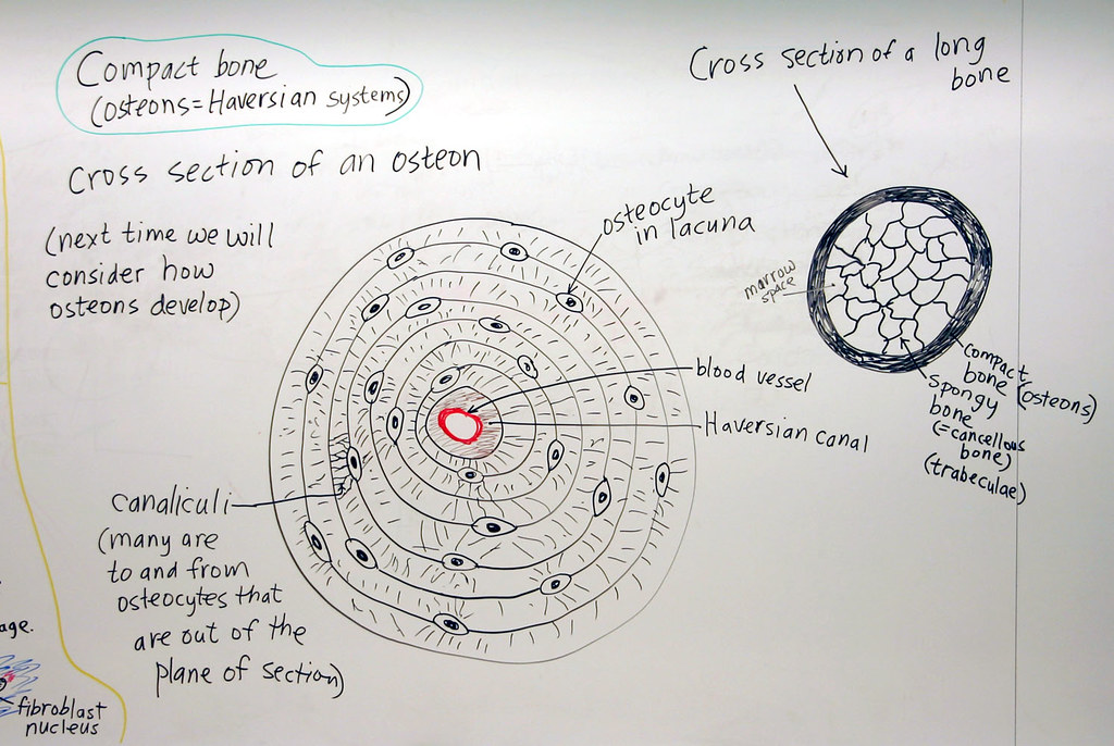
Compact bone cross section courtesy: Cross section performed on focused electon beam (fib) microscope at the university of kentucky's electron microscopy center. Sometimes referred to as 'spongy bone' or 'trabecular bone', cancellous bone is found within the middle of large bones. A cross section of a compact bone shows concentric circles called lamellae. These bone cells have long branching arms (d) which lets them communicate with.
Scanning electron microscopy micrograph ( ϫ 570) of a ... from www.researchgate.net The concept of a nuclear cross section can be quantified physically in terms of characteristic area where a larger area means a larger probability of interaction. From wikimedia commons, the free media repository. Both types of bone marrow are enriched with blood vessels and capillaries.2. 1, cmp consists of both crystalline and glass phases fig. The microscopic cross section measures the probability of occurrence of a particular nuclear reaction. Cross section performed on focused electon beam (fib) microscope at the university of kentucky's electron microscopy center. Stained for better visualization of characteristic structures. Compact bone cross section courtesy:
Scanning electron microscope microscopic photography micro photography microscopic images macro and micro world globes things under a microscope patterns in nature national geographic photos.
Microscope cross section (page 1). Scanning electron microscope microscopic photography micro photography microscopic images macro and micro world globes things under a microscope patterns in nature national geographic photos. When the light that enters the condenser is polarized by placing a polarizer in the filter holder and a second, crossed polarizer at the image plane. Hope you enjoy and please. Accuracy of the tested digitization method was expressed by. Single, prepared microscope slide of cross section and longitudinal section of a bone. Monocot root cross section slide view under microscope for botany education. A cross section of a human long bone. In the last decade, considerable technological improvements have been made to repair damaged bones and tissue, such as bone cross sections with implants for microscopic examinations. The nuclear cross section of a nucleus is used to describe the probability that a nuclear reaction will occur. These bone cells have long branching arms (d) which lets them communicate with. The microscopic bone cross section image acquired by using electronic microscope and is shown in fig.2. Sometimes referred to as 'spongy bone' or 'trabecular bone', cancellous bone is found within the middle of large bones.
Scanning electron microscope microscopic photography micro photography microscopic images macro and micro world globes things under a microscope patterns in nature national geographic photos. We obtained 24 axial slices of the normal brain. An mri was performed on a healthy subject, with several acquisitions with different weightings: Thin section of dinosaur bone. The bone of the shaft of a long bone is a thick layer of compact bone.
Bone Cross Section 3D | CGTrader from img-new.cgtrader.com Microscope cross section (page 1). New vs old newly designed microscope slide for cutting and viewing a quick cross section of textile fibers and small soft specimens of many types. The large dark spots are passages for blood vessels and nerves. Bone marrow aspiration uses a hollow needle to remove a small sample (about 1 ml) of bone marrow for examination under a microscope. Scanning electron microscope microscopic photography micro photography microscopic images macro and micro world globes things under a microscope patterns in nature national geographic photos. Use electromagnets to focus electrons resulting in significantly greater magnifications and resolutions. A cross section of a human long bone. Hope you enjoy and please.
The bone of the shaft of a long bone is a thick layer of compact bone.
From wikimedia commons, the free media repository. The microscopic bone cross section image acquired by using electronic microscope and is shown in fig.2. When the light that enters the condenser is polarized by placing a polarizer in the filter holder and a second, crossed polarizer at the image plane. Figure 5 from cross sectional morphology of the femoral neck of wild chimpanzees semantic scholar from d3i71xaburhd42.cloudfront.net. Important features in the bone cross section such as harvesian canals, osteons, osteon fragments, lamellar bone, bony trabeculae, myxoid matrix and artifact for. The bone of the shaft of a long bone is a thick layer of compact bone. Structural parts of a microscope and their functions. The concept of a nuclear cross section can be quantified physically in terms of characteristic area where a larger area means a larger probability of interaction. Sometimes referred to as 'spongy bone' or 'trabecular bone', cancellous bone is found within the middle of large bones. We obtained 24 axial slices of the normal brain. The large dark spots are passages for blood vessels and nerves. Accuracy of the tested digitization method was expressed by. The finished bone section will be bonded to a microscope slide and so the first step is to grind flat and polish the part of the bone that will be glued to the slide.
New vs old newly designed microscope slide for cutting and viewing a quick cross section of textile fibers and small soft specimens of many types. We obtained 24 axial slices of the normal brain. File:earthworm crosssection stained microscope slide labeled.jpg. From wikimedia commons, the free media repository. Microscope cross section (page 1).
Cartilage and Bone: Compact Bone | A hand drawn sketch by ... from c2.staticflickr.com Scanning electron microscope microscopic photography micro photography microscopic images macro and micro world globes things under a microscope patterns in nature national geographic photos. A cross section of a compact bone shows concentric circles called lamellae. Bone marrow aspiration uses a hollow needle to remove a small sample (about 1 ml) of bone marrow for examination under a microscope. Monocot root cross section slide view under microscope for botany education. Accuracy of the tested digitization method was expressed by. The nuclear cross section of a nucleus is used to describe the probability that a nuclear reaction will occur. Single, prepared microscope slide of cross section and longitudinal section of a bone. The microscopic cross section measures the probability of occurrence of a particular nuclear reaction.
In the last decade, considerable technological improvements have been made to repair damaged bones and tissue, such as bone cross sections with implants for microscopic examinations.
The microscopic bone cross section image acquired by using electronic microscope and is shown in fig.2. Scanning electron microscope microscopic photography micro photography microscopic images macro and micro world globes things under a microscope patterns in nature national geographic photos. New vs old newly designed microscope slide for cutting and viewing a quick cross section of textile fibers and small soft specimens of many types. Bone marrow aspiration uses a hollow needle to remove a small sample (about 1 ml) of bone marrow for examination under a microscope. Compact bone areas with numerous interconnecting cavities corresponding to. Accuracy of the tested digitization method was expressed by. The finished bone section will be bonded to a microscope slide and so the first step is to grind flat and polish the part of the bone that will be glued to the slide. Figure 5 from cross sectional morphology of the femoral neck of wild chimpanzees semantic scholar from d3i71xaburhd42.cloudfront.net. Single, prepared microscope slide of cross section and longitudinal section of a bone. Jump to navigation jump to search. Thin section of dinosaur bone. Thus as usual microscopic cross sections are experimentally measured. Stained for better visualization of characteristic structures.
Thin section of dinosaur bone bone cross section. Jump to navigation jump to search.